

For those individuals who have needed to undergo some sort of medical imaging test recently, like a CT scan, you probably have heard that there is a slight risk of radiation exposure. This is true of many medical imaging procedures, such as CT scans, X-rays, and nuclear imaging tests. Since the radiation exposure from medical imaging tests has become a common topic of recent in the medical media, patients have been increasingly concerned about the risks of radiation exposure. Specifically, patients are concerned whether the radiation exposure will increase their risk of developing cancer later on. Continue Reading Article >
Radiation Oncology: Understanding Different Treatment Plans

Cancer can be one of more complicated and difficult diseases people experience, and, while we may not have developed the perfect cancer treatment yet, cancer treatment has reached very advanced levels in accordance to the levels of complication with cancer.
One of the more common and effective ways to treat cancer is with radiation therapy.
The radiation used in treating cancer is ionizing radiation since it forms ions in the cells that the radiation passes though. Ions are electrically charged particles, so the radiation creates ions by removing electrons from the atoms and molecules inside the cells, which ends up killing the cells or altering the genes in a way that causes the cells to stop growing.
Although every radiation treatment uses ionizing radiation, it is important to remember that there are many different types of radiation treatments. The differences result from the different machinery, tools, execution, and techniques used when performing the therapy.
The differences between the different treatments are very significant as they lead to different energy levels in the radiation, which dictates have deeply and strongly the radiation will penetrate into the body’s tissues.
It is very important to have an idea what the different radiation treatments are because when it comes to planning a treatment, the radiation oncologist will work with you or your loved one to determine which type of radiation plan will be most suitable for your specific circumstances. So, here’s a quick guide to the various radiation treatments:
External Beam Radiations
External-beam radiation therapy is typically delivered using a machine called a linear accelerator (or a LINAC). By using electricity to form a stream of fast-moving subatomic particles, a LINAC creates high-energy radiation, which treats the cancer.
3D-CRT: 3-dimensional conformal radiation therapy, or 3D-CRT, utilizes highly-advanced computer software and treatment machines that allows doctors to deliver radiation more effectively by targeting areas that conform to very precisely formed shapes.
IMRT: Intensity-modulated radiation therapy, commonly known as IMRT, utilizes hundreds of collimators (tiny radiation beam-shaping tools) to deliver a single dose of radiation. IMRT specifically allows the doctors to modulate how high the level of radiation is being delivered to specific areas and then determining how many doses to use for each specific spot.
IGRT: Image-guided radiation therapy, or IGRT, utilizes repeated imaging scans, such as MRIs and CT scans, so the doctors can identify changes in the tumor’s size and location. This allows them to adjust the patient’s position, the radiation doses, and the duration of the treatment accordingly, which increases the precision of the treatment.
Tomotherapy: Tomotherapy is sort of a hybrid between IMRT and IGRT as the machine both delivers radiation, as an external-beam radiation therapy machine would, and also provides images of the patient’s tumor immediately before treatment. This increases the precision of the radiation and spares more normal tissue.
Stereotactic Radiosurgery: Stereotactic radiosurgery, or SRS, delivers one or more very high doses of radiation to a small tumor by utilizing extremely accurate image-guided targeting techniques and strategic patient positioning. However, to avoid causing excess damage to surrounding healthy tissue, SRS can only be used to treat small tumors with very well-defined edges.
Stereotactic Body Radiation Therapy: Stereotactic body radiation therapy, or SBRT, delivers its radiation therapy in fewer sessions than other treatments typically do. SBRT uses smaller radiation fields with much higher doses than treatments such as 3D-CRT. Again, SBRT is only used to treat small, isolated tumors, such as cancers in the lung and liver. Doctors usually refer to SBRT systems by their brand names, which is most commonly the CyberKnife.
Proton Therapy: While the previous radiation therapies utilized photon beams, this treatment uses proton beams to deliver charged particles to the cancerous area. While photon beam radiation deposits its energy semi-evenly along its way through tissue, proton beams deposit much of their energy at the end of their path and deposit less along the way. This hypothetically decreases the amount of radiation being exposed to healthy tissue and increases the amount of radiation being exposed directly to the cancer.
Other Forms of Radiations
Internal Beam Radiation (Brachytherapy): Brachytherapy differs from the external beam radiation treatments as it utilizes radiation from a radiation source being placed inside or on the body. The sources usually involve radioactive isotopes being sealed in tiny pellets or seeds, which are then placed in patients using tools such as needles, catheters, and other forms of delivery techniques. Then, the isotope naturally breaks down, which results in the release of radiation that likely damages nearby cancer cells.
System Radiation Therapy: System radiation therapy involves the intake or injection of a radioactive substances, such as radioactive iodine or a radioactive substance bound to a monoclonal antibody. The iodine is used in cases with thyroid cancer as the thyroid naturally takes in the iodine substance, and other cases use the substance bound to the monoclonal antibody as the antibody naturally travels to and targets the infected cells where the tumor is located.
At the end of the day, there are many, many different ways to treat cancer. So, when it comes to planning your treatment, you can prepare yourself or your loved ones by just becoming familiar with some of the different treatments and how they work, that way you can be on the same page with your radiation oncologist. Also, always remember that while all of this information can be overwhelming, you can always ask someone on your radiology team or your health care provider any questions you may have, as they can likely provide you with the right answer.
By Russell McBurnie
Do I Need a CT Scan or an MRI?
Diagnostic imaging allows doctors to examine inside a patient’s body to gather clues as to what may be causing the patient’s complications. Of course, there are many forms of diagnostic imaging tests depending on many factors. The common types of tests include X-ray, CT scan (also known as CAT scan), and MRI scan. Continue Reading Article >
Finding Diagnostic Imaging Prices in Texas

Whether it’s to determine if your ankle is sprained or broken, or to see what’s causing pain in your chest, sometimes doctors use diagnostic imaging to examine inside a patient’s body. Depending on certain factors such as the area and the symptoms, there’s a variety of machines and techniques the doctor can use to examine a patient.
The different types of tests include common examination procedures most people are familiar with, such as ultrasounds, X-rays, and MRIs, but there’s many other types as well. Since most diagnostic imaging experiences are quick, easy, and painless, people aren’t typically concerned with undergoing the test itself.
Rather, individuals are often concerned about what type of damage will be inflicted on their bank account as not all tests are covered by health insurance for a number of reasons and circumstances. Since people don’t like finding out that they owe thousands of dollars after the fact when they get a letter in the mail, finding prices for diagnostic imaging is very important to do beforehand.
Of course, finding prices is easier said than done. Many people don’t even know where to start as providers don’t really have a price menu on their websites. Luckily, there are tools out there that can help you find the best price available for you.
Save On Medical
They say everything’s bigger in Texas, but that certainly does not need to be true when it comes to your medical expenses, especially with the help of Save on Medical.
Save On Medical is a tool that helps people find prices for diagnostic imaging tests, among other procedures and medical-related things. Their website helps you find which provider is best for your needs by comparing all of the prices, quality, and convenience for each provider. Then, if you decide to book your procedure on their website, you’ll lock on to their discounted price.
Moreover, Save On Medical also allows people to schedule their appointments, manage them, pay for them, read about other people’s experience with a certain provider, and write their own reviews about their experiences with a provider. Overall, they offer a great tool when it comes to finding not only the prices for diagnostic imaging, but overall finding the best provider.
Finding Prices Using Save On Medical
Save On Medical makes using their tools very easy. So, for those who navigate the internet regularly should have no trouble figuring out how to find the princes. But, for those who aren’t internet-savvy to any degree, so here’s a quick guide how to find prices.
1. Select the dropdown menu titled “Find Radiology” on the top of their home page, and select your desired procedure. The list includes many different types of diagnostic imaging, and each type of test is categorized by which area of your body is being examined. The types of diagnostic imaging include:
· Angiography
· CT
· Fluoro
· MRI
· Mammography
· X-Ray
· And, other.
2. Enter the location for the procedure. The box is directly next to the first menu, and you can enter the location in the form of “city, state” such as Dallas, Texas, or you just use the zip code (75204, for instance).
3. Finally, Save On your medical expenses and book your appointment! Once the results appear, you can then easily compare them and search which provider offers the prices that best match your needs.
At the end of the day, hopefully you discover that not everything in Texas needs to be bigger. After all, medical bills are already big enough. And, in addition to any price tools online, don’t be scared to ask your doctor about where you should go for an examination. Your doctor might be able to provide you with the best recommendation as they are experts that understand your individual circumstances.
By Russell McBurnie
Finding diagnostic imaging prices in Georgia

When doctors need to take a look inside of your body to examine medical complications and issues, they use diagnostic imaging. Diagnostic imaging tests are performed using different machines and various techniques depending on a few factors such as the area of the body that’s being affected and the symptoms that the patient is experiencing.
Various types of diagnostic imaging are used by doctors, but the common forms include tests such as MRI scans, CT scans, and X-Rays. The procedures are usually very easy, quick, and painless, but some may involve laying down in a certain position for an extended amount of time. Less common forms of diagnostic imaging require anesthesia, especially if they involve the use of a scope.
Most diagnostic imaging, however, is easy and painless. So, patients are not typically concerned with the test itself. Instead, patients are concerned about other things. One concern for many patients is how much it will cost. Not all of the tests are covered under health insurance, or they’re only partially covered.
Whether it’s because of deductibles, monthly limits, or some other restriction, it can end up costing the patient a lot of money. So, for those individuals, how do you ensure that you are paying the best price available without finding out when you first get the bill in the mail? Well, luckily, there’s a pricing tool that makes it very simple!
Save On Medical
By allowing you to search for a procedure and then compare the prices, quality, and convenience of the various providers, Save On Medical is the perfect tool to help you find prices for diagnostic imaging. Additionally, you can also schedule and manage your appointments, pay for your procedure, and read and write reviews about your experiences with certain providers.
Not only is a very useful tool to find pricing, but it also helps you save on your costs as you can lock in discounted prices by booking your procedure through their website. So, if you are looking for the best deal on an MRI, X-Ray, or some other diagnostic imaging test, Save On Medical is the perfect tool for you.
Finding Prices with Save On Medical
Like everything else on the interest these days, navigating the Save On Medical website is very simple. But, in the case that you can’t figure it out for a certain reason, here’s a quick three step guide to finding your prices:
1. First, find the dropdown menu toward the top of their home page and select the specific type of procedure you are searching for. The procedures are categorized by the type of diagnostic imaging test and then by which area of the body you would like to examine with the test. The types of tests available are:
· Angiography scans
· CT scans
· Fluoro scans
· MRI scans
· Mammography scans
· X-Ray scans
· And, other.
2. After you select the procedure, you need to enter in the location you would like to search in right in the box next to the procedure box. Just enter either the city, state or enter in the zip code. So, for example, type “Atlanta, Georgia” or “30023” for the zip code.
3. Then, after you click the “search” button, you can compare all of the prices, find the one that’s right for you, and book your appointment! It really is that easy. It’s kind of like looking for flights, just type in what you need and where, and see if any of the results work well for you.
At the end of the day, there’s no need to overpay for diagnostic imaging, especially since the tests can already be pretty pricy. Just remember that there’s always more to your decision than just the prices, so make sure that you’re looking for a provider that offers adequate quality, convenient appointments, and a good price! And, if you have any questions about the providers, tests, and what’s right for you, never hesitate to ask your doctor as your doctor is an expert that might better understand your individual circumstances.
By Russell McBurnie
Magnetic Resonance Imaging Prices in Florida
Magnetic resonance imaging allows doctors to search for medical issues or certain conditions within a patient’s body. Medical imaging allows patients to quickly find a cause to their problems. If you're wondering, "How much does an MRI cost in Florida?" we've got your back. Continue Reading Article >
Finding Diagnostic Imaging Prices in Alabama

Diagnostic imaging provides doctors with the ability to look for complications and issues within a patient’s body. There are many different machines and techniques when it comes to diagnostic imaging, and the procedure depends on the symptoms of the complication as well as which area of the body is being affected.
The various types of diagnostic imaging that doctors typically use to test for medical issues include tests like X-rays, CT scans, and MRIs among many other. Since most of the tests involve easy, painless, and simple procedures, people don’t usually stress about the test itself.
Rather, patients stress about the costs of these tests as they can go well into the thousands and they’re not always covered fully or at all under health insurance. So, whether you need to pay deductibles, you’ve reached your monthly limit, or it’s simply not covered, you probably want to know how much diagnostic imaging costs.
But, how are you supposed to figure that out? Then, how are you supposed to find the best priced procedure? Well, luckily, the answer is a lot easier than you might’ve thought.
Save On Medical
Luckily, if you have any concerns regarding the costs of diagnostic imaging tests and procedures, Save On Medical is an amazing tool that makes that task extremely easy.
Save On Medical allows people to search different procedures and then compare the costs, quality, and convenience of each provider that offers such procedures in your area. Then, by booking on the website, you lock in the discounted price offered by Save On Medical.
In addition to comparing costs, Save On Medical also allows people to make appointments, manage their appointments, pay for their procedures, read about other people’s experience with certain providers, and write their own review about their experience with certain providers.
How Do You Find Prices Using Save On Medical?
As most websites on the internet today, Save On Medical makes using their tools very easy. But, not everyone is internet-savvy to any degree, so here’s a simple guide how to find your prices.
1. Once you are on their home page, select the dropdown menu titled “Find Radiology” and select your desired procedure. The list includes many different types of diagnostic imaging. Each type is then broken down by which area of your body you will need examined. The types include:
· Angiography
· CT
· Fluoro
· MRI
· Mammography
· X-Ray
· And, other.
2. Enter the location you wish to find the procedure around. You can enter it in the form of “city, state” such as Birmingham, Alabama, or you can enter in the zip code (35208, for instance).
3. Finally, Save On your medical expenses! Compare the given results to search which price and provider best matches your needs, and then you can schedule an appointment and undergo your procedure.
At the end of the day, finding a decent price for your procedure might not be as difficult as your first thought, which is good since you could be spending quite a bit when it comes to diagnostic imaging. In addition to Save On Medical, or any other price tools, don’t be scared to ask your doctor about where you should go for your test. Your doctor might be able to provide you with the best recommendation as they are experts that understand your individual circumstances.
By Russell McBurnie
3D Ultrasound: Myths Debunked
3D Ultrasound was developed in 1987 at Duke University. It was discovered by Olaf Von Ramm and Stephen Smith. It is primarily used to look at the development of the fetus in pregnant women and detect birth defects and other problems. Parents learn the sex of their child and about serious medical conditions or birth defects. It is also used for screening for breast cancer and other illnesses. Continue Reading Article >
10 Questions for your Radiation Oncology Team

While cancer will always be a difficult experience, there are certain things you can do to make it a little less stressful. One thing that will almost certainly help you decrease some stress is adequately preparing to better understand the experience as best as you can.
One way to prepare and understand the experience is to ask your radiation oncologist certain questions you will need to know the answers to. This way you will better understand what to expect during your battle. To help you prepare, we’ve listed 10 questions you might want to ask your radiation oncology team:
1. Do you recommend radiation therapy to treat my cancer?
2. What type of radiation therapy should I receive to best treat my specific cancer considering my specific circumstances?
3. What are the benefits of undergoing radiation therapy, and do they outweigh the effects of radiation?
4. What are the side effects of radiotherapy?
5. How many treatments will I need to undergo, and how long does each session last for?
6. What are the different stages of cancer I should expect, and what are the cancer survival rates for cases similar to mine?
7. How do I best manage the side effects of radiations?
8. What should I expect as the normal level of skin irritation and fatigue that I’ll experience during my treatment?
9. What are the potential long term side effects of my treatment, and how do I minimize those effects?
10. Where will I undergo my treatment and where will I meet with the medical professionals during my treatment?
At the end of the day, it is important to remember that each member of your radiation oncology team is there to assist you. So, try your best not to get stressed about what’s going on as they are always there to help. And, of course, always keep in mind that they will be there for when your questions arise, so don’t feel as if you need to ask your radiation oncologist all of your questions during the first meeting.
By Russell McBurnie
6 Celebrities Who Promote Mammograms

Almost everyone has some type of celebrity that influences them in some way or another. And no, not every celebrity influence has to be something bad or pop culture related. Celebrities can, and often do, influence good things just as often as not.
For some people, celebrities with breast cancer are extremely influential, especially when it comes to screening. Getting a mammogram is crucial when it comes to detecting breast cancer early, and an early detection makes the battle against cancer a lot more manageable.
( Find Mammogram Cost Resources at www.saveonmedical.com )
Despite the importance of screenings and early detections, women don’t always stay on top of getting their mammograms. Luckily, however, there are celebrities that have to influenced women (and continue to do so) to make sure they are getting screened. Here are 6 of those celebrities:

1. Sheryl Crow –
Sheryl Crow, best known for her country and folk music, was diagnosed with breast cancer in 2006. Without any prior symptoms of breast cancer, her mammogram was crucial for getting her cancer detected before it grew to an extreme level. She now encourages other women to make sure they keep up with their screenings, as it can be the key to getting an early detection.

2. Sandra Lee –
Sandra Lee, best known as a celebrity chef and an author, was diagnosed with cancer when she was 48 years old. She also had no clear symptoms of having breast cancer, but came to a diagnosis because of her routine mammogram. Because of that, she now encourages others to do the same and even went on Good Morning America to do so.

3. Rosie O’Donnell –
Best known as a comedian, actress, and TV personality, Rosie O’Donnell often encourages other women to keep up on their routine screenings so they can prevent a difficult battle with breast cancer. She explained on her show that she had a breast cancer scare after her first mammogram, where she had two spots, one in each breast, that might have been breast cancer. Fortunately, they were both benign, but since her scare she has encouraged that other women stay on top of their mammograms.

4. Amy Robach –
The co-host of the Today Show received a mammogram on live TV in 2013. Having avoided her routine screenings for the years leading up to her live mammogram, she was shocked to find out that her results came back positive for breast cancer leading her to encourage other women to take the precautions that she had avoided.

5. Nancy Reagan –
Ronald Reagan was not the only influential Reagan, as the former First Lady of the United States, and former actress, was also extremely influential. After being diagnosed with breast cancer in 1987, while her husband was still the president, she influenced many people to undergo the proper guidelines for mammograms as she was able to successfully remove her cancer due to her detection.

6. Andrea Mitchell –
The chief foreign affairs correspondent on NBC was diagnosed with breast cancer in 2011 because of her routine mammogram. Her cancer was detected early enough to prevent the cancer from spreading anywhere, and she now encourages others to take the same precautions that helped her protect herself.
At the end of the day, it doesn’t matter where your influence comes from so long as you make sure you take the proper precautions to care for yourself by preventing a difficult battle with cancer or cancer altogether. But, for those who find their inspiration from their favorite celebrities, there are plenty who promote mammograms.
By Russell McBurnie
Mammograms: They’re not just for women!

When most people hear about breast cancer, the negative diagnosis is usually associated with women, but men are susceptible to breast cancer as well. Breast cancer in men is infrequent, but it can transpire. Statistics show that about one percent of all breast cancer cases occur in men. Typically, a mammogram is a tool used to help identify cancer in men and women.
Mammograms
Initially, a mammogram is an X-ray image that is taken of your breast and used to screen for breast cancer. Mammograms can often detect breast cancer in its early stages, which in turn lowers the risk of the cancer developing and can often save lives due to its early detection. During the procedure, your breasts will be pressed between two sturdy surfaces and the breast tissue will be spread apart, so that the doctor can take the X-ray and then examine the pictures for signs of cancer.
Breast Cancer signs in Men
Men will often contain far less breast tissue as opposed to women. Often times, the cancerous signs are easier to spot in men than in women. A prevalent sign of breast cancer in men is a painless lump in the chest region. Sometimes it may even be a thickening in the chest, breast or underarm area. Mammograms are great for identifying lumps long before they can be touched or seen.
If you notice any alteration in the nipple like it is retracting, itching, or possibly even a scaly rash of the nipple, it may be Paget's disease, which can be linked with breast cancer. Paget disease is an uncommon cancer on the nipple. It has a darker circle around it called the ‘areola.’ While the majority of Paget cases occur in women, it also is also a cancer that men should keep an eye out for. Some more common examples for men with possible breast cancer symptoms are a dripping from the nipple. This is often caused by a blood soaked inflammation of the breast, or you might see a wound in the skin of the breast.
What should you do if you have Breast Cancer?
Men can often become prideful in regards to their health. With a possible case of breast cancer, it is not different. However, if you are a man or woman and you notice a change in your breast or chest area, you should contact a healthcare provider as soon as possible. The sooner you do this, the better. Breast cancer offers the highest survival rate when it is discovered earlier. A good way to find a competent and trustworthy healthcare provider is to receive a referral from a family member or friend. If you can’t do that, then call your local clinic, health department or hospital.
By Preston Copeland
Do I Need an X-ray of my Ankle?

At one point or another, most people will experience some type of a possible ankle injury during their lifetime. Whether you just twisted your ankle on the stairs, rolled it off of the sidewalk, or slammed it while you were playing your favorite sport, ankle injuries can hurt a lot.
Of course, depending on how serious the injury is, there are different ways to go about how you handle treating your injury. Maybe you just need to put an icepack on it for a little, or maybe you can just walk it off, but if you’re thinking, “is my foot broken?” you might need an X-ray.
In order to detect a fracture, or a broken bone, you will need to get an X-ray. However, this can be harder to determine than many people imagine. So, when you ask yourself, “do I need an X-ray?” there are certain things you should be looking for. Here are some tips to help you figure out whether you should get an x-ray –
At First Glance:
Unsurprisingly, there are some ankle injuries that are clearly serious enough for an x-ray and some that are a lot less obvious. So the first step in determining whether you need an x-ray or not will be to look at the ankle that is hurt and compare to the ankle that isn’t after it happens.
If there are significant differences, such as a deformed bone, or severe swelling and bruising, you will probably want to get an x-ray immediately. However, if there is no obvious deformity, and your ankle definitely hurts, it can be a lot more difficult to decide whether you need an x-ray.
Ottawa Ankle Rules:
Luckily, there are specific guidelines that medical professionals typically use to determine whether a patient will need an x-ray called the Ottawa Ankle Rules, and you can follow these guidelines to help you decide how serious you believe your ankle injury might be. So, here are the guidelines –
You will probably need to get an x-ray if:
1. You experience a significant amount of pain on the outside of your foot where your little toe is, specifically around the fifth metatarsal as well as on top of your foot by the navicular bone, which is on the top of your foot close to where the heel is.
2. You feel a lot of pain when you or someone else touches the back half of the malleolus, which is the ball looking thing on the side of your ankle, or six centimeters above and/or below the malleolus.
3. Or, you experience extreme pain or the inability to walk four steps immediately after the injury and at the time of examination.
You can always use these guidelines to determine whether you will need an x-ray in the case that there is no obvious or significant deformity to the ankle soon after the injury occurs.
At the end of the day, if you are worried that you may have broken a bone in your ankle, then a trip to the doctors won’t hurt (unless, of course, you walk there). A doctor, or an orthopedic expert, can always give you educated advice on how to properly care for your injured ankle.
By Russell McBurnie
Am I Old Enough for a Mammogram?
For the past few decades, women and researchers have been trying to figure out when women should start getting their annual mammogram or 3D mammogram. A diagnostic mammogram screens for breast cancer, and it is critical in order to detect cancer as early as possible. Early detection is one of the best tactics in effectively beating cancer, so this topic of the utmost importance. Continue Reading Article >
10 Reasons You Might Need An Ultrasound

An ultrasound is a machine that uses high frequency sound waves to capture images in the body. This is so doctors can make a diagnosis. It is similar to radar which helps the military detect planes and ships. It lets the doctor see your organs, tissue, and blood vessels before making an incision. The results help in getting a correct diagnosis. Fortunately, this useful procedure uses no radiation. So, you be questioning, "Why do I need an ultrasound?" Here's 10 reasons you might end up undergoing an ultrasound.
Pregnancy
An ultrasound is often used to track the progression of a pregnancy. It is used to make sure that all is going well and to catch problems before they occur. It shows what the baby looks like, the size, and how the organs are functioning. When complications arise often an ultrasound can detect the problem. They determine the due date, birth defects, multiple pregnancies, and position of the baby.
Gynecology
It is used to detect tumors and tissue growth in the vagina, uterus, Fallopian tubes, and ovaries. Often this test can detect abnormalities of the tissue so the doctor can diagnose and treat serious condition that affect women. It is used for fibroid tumors, endometriosis, ovarian cysts, and uterine growths.
Heart Problems
An ultrasound is used to diagnose problems with the heart. It can determine if a heart valve is leaking or not working correctly. It measures the heartbeat for irregularities. The test can even detect blood clots in the heart and infections!
Blood Vessels
This test is used to detect blood clots in veins. It is used on stroke patients to see if main arteries are blocked. The test can often tell why veins are swollen due to a blood clots or deep vein thrombosis.
Abdominal Structure
The ultrasound or 3D ultrasound can be used to access most solid organ in the body. This means the gallbladder, kidneys, pancreas, bladder, prostrate, uterus, and ovaries. It can test for gallstones or an infected gallbladder.
Neck
It is used to detect problems with the thyroid in the neck. The test is used to detect nodules, growth, and tumors.
Knee Joints
It can be used to detect fluid in knee joints from injuries and cysts that grow there.
Guide Needles into the Body
Sometimes an ultrasound is used to guide needles into the body. When an intravenous line is needed but is difficult to find a vein this test is used. Often this is used when the needle must go into the neck, groin, or chest where injections are more difficult to see or administer.
Prostrate and Urology
Often an ultrasound is used to look at prostate glands in men and to examine the rectum. It is often used to measure the blood flow of the kidneys, detect kidney stones, and early prostate cancer.
Biopsies
It can be used by a surgeon to take a biopsy of a tumor that may be cancerous. It helps them find the locations so that they can take the biopsy with accuracy.
You may need an ultrasound when your doctor wants to make a diagnosis and confirm results before proceeding with treatment. It is an effective test in determining many different disease, tumors and complications in the body. A 3D ultrasound will show pictures of your organs or baby in 3 dimensional formats. These images will be moving. An ultrasound is a painless test that takes ½ hour to 45 minutes.
By Joan Russell
7 Reasons You Might Need An X-Ray

For all of you out there this blog should be especially important to you. The summer gives all of us a little extra time to check off those projects we promised ourselves we would do. Before getting into full-out DIY diva mode and taking on potentially dangerous tasks though, you should first ask yourself “Is this a good idea?”
If the answer is yes, ask yourself, “Is this safe?” If you get a yes there you are good to go, and good luck. If any of these questions yielded a no, and you still felt inclined to accomplish “said task” and somehow managed to injure yourself or anyone else around you… read below and answer this final question, “Do I need an X-Ray?”
It’s not always easy to tell whether or not you need an x-ray, but there are a few situations that you may find yourself in this summer where there’s a pretty good chance you might. Here are seven situations that may be an indicator that you need to head to the doctor’s office for an x-ray:
1. You’ve fallen and you can’t get up.
If you attempted to clean out the copious amounts of leaves summer showers left behind in the gutters on your roof, and the ladder failed you…you may need an x-ray.
2. You missed that invisible step and fell flat on your face.
Whether you're simply leaving a store and didn't see the caution "Watch Your Step" sign or lost your footing while walking down the stairs, you might end up with a sprain. Don't worry, it happens to the best of us.
3. You’ve got a Warriors fan and a Cavs fan under the same roof.
Basketball and hockey are both finishing up their seasons. If you live in a divided household, the race to the remote can be dangerous. You won, but your pinky is now twisted a strange direction.
4. Your synchronized diving took a turn for the worse.
Let’s say while performing your latest diving trick into the pool, your finish had a different outcome than you’re used to. The pain has been consistent for three days now, and you’re pretty sure that it’s not just your pride.
5. Your idea of fun and games end up being not so fun.
An intense game of beach kadima resulted in a loss of footing and perhaps a twisted ankle.
6. Your dumbbells are out to get you.
While training for your beach bod in May you dropped a dumbbell on your toe. You managed to walk it off, but it’s now almost June and your toes seem to be doing something funky…looks like there won’t be any Baywatch in your future.
7. Your toddler took your game of dice to a whole new level.
One second you’re sitting at the table rolling dice with your toddler, the next minute two dice has turned into one, and Tommy is giving you a devilish grin.
If any of these scenarios or something similar have happened to you this summer, its safe to say…you might need an x-Ray.
By Maren Burns
The Many Uses Of X-Ray
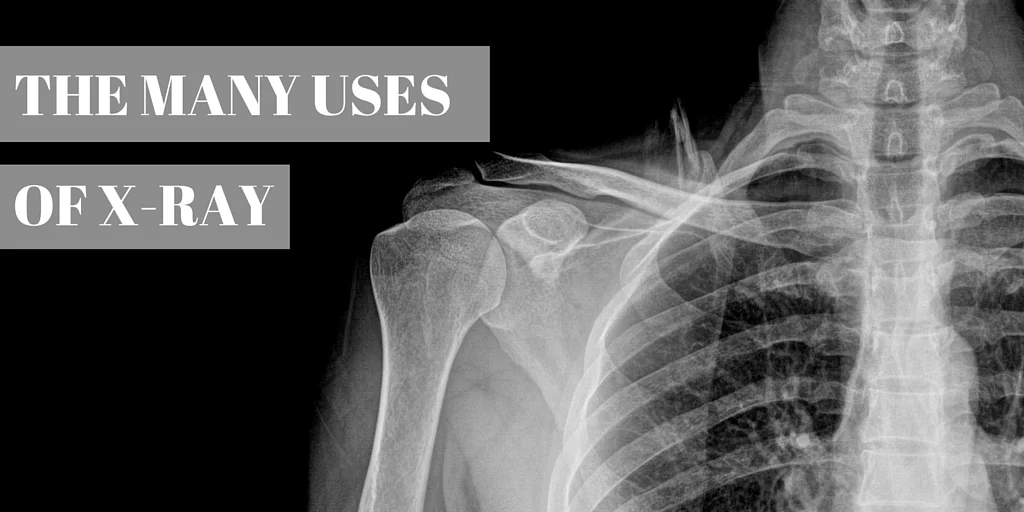
What is an X-Ray? An X-Ray is “an electromagnetic wave of high energy and very short wavelength, which is able to pass through many materials opaque to light” At some point in your life you’ll probably find yourself asked the question, “Do I need an X-Ray?”
For many, the answer is yes. Don’t be alarmed, though! The process is quick and painless, and the amount of radiation you’re exposed to during the procedure is harmless to adults. This radiation, however, is not safe for a developing fetus. Be sure to tell your doctor about any questions or concerns prior to the procedure, especially if you’re pregnant.
An X-Ray is performed for three main reasons:
1. To photograph an area of the body experiencing pain
2. To record the progression of a disease
3. To monitor the effect of a treatment
But there are many additional instances in which an X-Ray can be helpful to a doctor.
Bones & Teeth:
Fractions and infections in the bones and teeth show up clearly on an X-Ray. For dentists X-Rays are helpful when trying to measure the amount of dental decay in a patient’s mouth. Also for victims of arthritis, X-Rays of the joints will reveal the extent of the disease to the doctor.
Chest:
The chest contains many important organs, and because of this chest x-rays make up a large percentage of all X-Rays taken. Mammography is an X-Ray procedure done to examine breast tissue and prevent breast cancer, while other X-rays of the chest can reveal lung and heart conditions as well. In some cases a contrast material will be injected into the body so that doctors can get a better look at where your circulatory system is experiencing problems.
Abdomen:
While primarily for digestive tract problems, X-Rays of the abdomen can also reveal to doctors the location of swallowed items. For example, if your child swallowed a key or a marble, an X-Ray of the abdomen would reveal the location of this item.
Written by: Maren Burns
Understanding The Difference Between MRI and CT
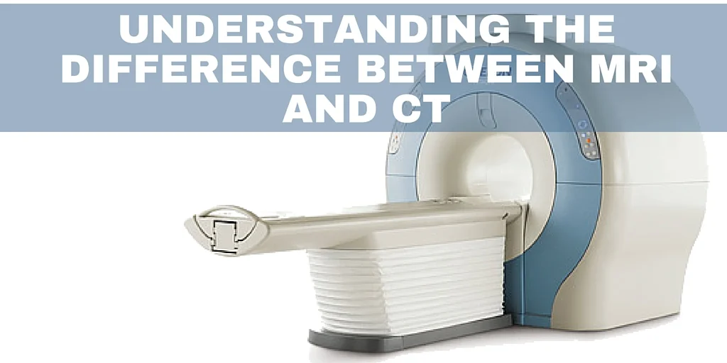
It definitely is not uncommon to walk into a doctor’s office for some sort of an appointment and not really understand what is going on. This is especially true in regards to undergoing specific procedures, especially if their functions tend to somewhat overlap. For instance, unless you clearly broke your leg and need an X-Ray, diagnosing a leg injury can be difficult. Maybe you cracked your femur, maybe you tore your ACL, or maybe you strained something. In order to figure out exactly what happened and what you need to do to better the injury, you must undergo certain procedures to see what happened to your body.
In such cases, as well as many others, the patient will undergo either an MRI or a CT scan. These procedures seem very similar on the surface, especially since they look almost identical. There is no need to ask why someone might not know the difference between the two, as they are very commonly confused. However, the difference is critical in determining the injury and the best means to recovery. So, it is important to understand what sets apart MRIs and CT scans. Let's take a look by first examining what exactly MRIs and CT scans are, and then how they are different.
MRI scans utilize very powerful magnetic fields and high-frequency radio waves in order to provide detailed images of various things inside a patient’s body. MRIs provide such detailed pictures by reading energy that is produced by water molecules after they re-align themselves following each pulse of radio waves.
What is a CT scan?
Unlike MRI scans, which use magnetic fields, CT scans utilize X-rays to provide details inside a patient’s body. CT scanners send X-ray beams through the patient’s body to the X-ray detector on the other side as the body moves through an arc-looking structure. While doing so, the CT scanner takes many pictures of what’s going on inside the body by comparing the strength of the beams, which are measured at about 1000 times per second.
Key Differences:
Primary Focus- One of the most significant differences between MRIs and CT scans is what they are typically used for, which, of course, is usually the determining factor for whether a patient would undergo an MRI or a CT scan, which is why understanding how a CT scan works versus an MRI is so crucial. MRIs provide very detailed images for soft tissue injuries due to how they function, so patient’s generally get MRIs for possible soft tissue injuries such as ones involving ligaments, tendons, spinal cord, brain tumors, etc. MRIs, however, cannot detect details regarding bone injuries because the lack of water in bones results in a black image where the bone is. On the other hand, CT scans are usually better for bone injuries, lung imaging, chest imaging, and cancer detection. So, depending on what the primary focus of your possible diagnosis, MRIs and CT scans might be of different help to you.
Accessibility- CT scans are more widely available than MRIs, but MRIs are also readily available to patient's just about anywhere. The difference in their accessibility might come down to price for some patients. A CT scan costs anywhere between $1500 and $3000, whereas an MRI can cost just over $4000 in some institutions. However, there is a reason for the price difference. CT scans are generally considered to be slightly more limited than MRIs in instances where the primary focus of the procedure does not distinguish between which machine would be better. Using X-ray to capture images, CT scans struggle to capture multiple, layered angles for more comprehensive imaging. So, often times when various angles are needed, patients receiving a CT scan must be repositioned. Contrarily, MRIs produce images using nearly every plane involved. While CT scanners might cause certain limitations regarding angles, MRIs cause certain limitations concerning implants. Patients with cardiac pacemakers, tattoos, and metal implants are sometimes unable to have the procedure due to the magnetism, whereas CT scanners can accommodate those patients.
Comfortability- While MRIs tend to be more versatile for the patient's needs, CT scans are definitely slightly advantaged in one category: patient comfortability. CT scans take a considerably shorter time than MRIs as they take about 5-10 minutes, including preparation time. MRIs, on the other hand, take between 30 and 90 minutes to capture the images alone. During that time, MRI patients are expected to remain still while the noisy machine performs its imaging. Many MRI patients could attest to the general discomfort, but it is not overwhelming. And, although CT scans are inarguably shorter and more comfortable, they are limited by size so heavier patients might prefer an MRI, especially when the patient's weight surpasses the 375-400-pound range.
Radiation Exposure- One of the significant differences between MRIs and CT scans that people typically find substantial is the difference in radiation exposure. While MRIs emit no radiation whatsoever, CT scans certainly do. CT scans emit an effective radiation dose of anywhere between 2 and 10 mSv. Such an amount is similar to the background radiation an average person is exposed to after 5 years of normal activity. Because of this, certain individuals, such as pregnant women, are very strongly advised not to undergo CT scans.
At the end of the day, your doctor is there to help you understand these differences as they are absolutely integral to following the best path for your injuries and issues. MRIs aren't better than CT scans and vice versa, they're simply just different procedures. However, doctors do not initiate conversations and discussions about whatever injury with patient's. Such a responsibility is on the patient, which is why we choose to go to the doctor's office. So, whether you want to know for the sake of knowing it or you actually want to know because you have a sore need and want to know the best approach moving forward, there is a critical difference between MRIs and CT scans. But, if you are not sure which procedure is best do not panic – your doctor should always be there to help you make sure you are taking the best course for whatever struggle you may be facing. So talk with your doctor about whether an MRI or a CT scan would be better for you and the conditions of your issue.
Written by: Russell McBurnie
Becoming A Radiologist: How Your Dr. Got Here

Your radiologist is the physician specifically trained to be the expert in medical imaging. Radiologists are doctors who specialize in diagnosing and treating diseases and injuries using various imaging techniques such as x-rays, computed tomography (CT), magnetic resonance imaging (MRI), nuclear medicine, positron emission tomography (PET) and ultrasound.
Interventional Radiology (IR), is a sub-specialty of radiology which uses image-guided minimally invasive techniques to diagnose and treat a large variety of medical conditions. IR can be an alternative to some surgical treatments such as epidural steroid injections or some oncology procedures.
Training requirements
From the very beginning, radiologists follow the same education path as every doctor in the U.S. They attend four years of undergraduate college and then four years at an accredited medical school, earning either a Doctor of Medicine (M.D) or Doctor of Osteopathic Medicine (D.O). They also complete an internship and residency in diagnostic radiology, and some physicians go on to complete fellowships in advanced radiology specialties such as vascular and interventional radiology, neuroradiology or mammography.
What role does a radiologist play in my health care?
Generally the provider taking your mammogram or x-ray is not the doctor, but a highly-trained radiologic technician. In fact, you may never actually meet the radiologist who reads your mammogram or MRI after the image is taken. Your radiologist is available to you and your referring provider by request, to ensure the correct study is ordered, to assist in the interpretation of the results and to discuss further examinations or potential treatments needed. Of course, during interventional procedures you will meet with the radiologist for your procedure.
After analyzing your images, the radiologist sends a report (an electronic written review) to your referring provider. Most of the communication regarding your exam goes from your radiologist straight back to your referring provider who will deliver your imaging results and formulate a management plan.
In general, a radiologist:
- Acts as an expert imaging consultant and provides a detailed report to the physician who ordered your exam
- Works with your referring physician to determine the appropriate imaging exam for your needs
- Reviews and interprets the images from your exam
- Recommends further exams or treatments when necessary and confers with referring physician
Key Skills and Strengths
Radiologists tend to be very analytical, detail oriented, and may have a strong interest in physics and technology. Knowledge in physics helps the radiologists with the proper positioning of the patient to ensure a clear image. Additionally, to become a successful radiologist, they must become experts at diagnosing and examining diseases using radiation or magnetic machines.
Certification and Accreditation
To ensure quality and safety, radiologists should also become board certified by the American Board of Radiology (ACR), a professional medical society dedicated to serving patients and society by empowering radiology professionals to advance the practice, science and professions of radiological care.
Written by: Marysa Stevens
10 Things To Look For In An Imaging Center
When your doctor decides it’s time to get a clearer picture of what’s causing your stomach pain, or you’ve hit 40 and need to schedule your first mammogram, you will be referred for diagnostic imaging. Just like when you’re looking for a dentist or a primary care provider, you want to do your research and find the best clinic for you. There are a few different things that make a great imaging center—here’s what to look for.
1. Medical team and expertise
Before you schedule your appointment or go to your exam, take a look at the medical imaging clinic’s website and get to know the business. Most likely, you don’t interact with the radiologist (your exam is performed by trained radiologic technologists, and the radiologists analyze your exam on screen in a separate dark room), but you still want to be comfortable knowing the doctor reviewing your exam is qualified and experienced. Most websites offer bios of their doctors and their medical team, look for specialty training and certifications.
2. The most appropriate exam
Remember, this is diagnostic imaging. The pictures are to help doctors understand what is causing you pain or illness. The radiologist works with your primary care provider or the specialist who referred you for imaging to make sure you’re getting the right exam, saving you time, money and potentially unnecessary radiation exposure.
3. State-of-the-art technology
Much like the most advanced smart phone or TV, technology in medicine is always changing especially in imaging technology. Mammograms can now be viewed by the radiologist in 3-D, meaning the breast tissue is more dimensional, in slices, allowing for a more accurate read. This eliminates false positives and also reduces anxiety for the patient. MRIs are also seeing improvements, with more power for cleaner reads and a wide-bore, or open MRI to enhance patient comfort.
4. Somewhere that won’t break the bank
Shop around for clinics that offer the most affordable exams and always make sure that your exam and the clinic is covered by your insurance provider.
5. Scheduling ease and availability
Nothing is worse than waiting to find the right answers. If you call to schedule your appointment but can’t get in for two weeks, you should look for another clinic. Most top imaging centers try to get their patients in for diagnostic imaging within 2 – 5 business days.
6. Customer service and friendly staff
Outpatient imaging centers have a very large staff who you interact with: schedulers, receptionists, radiologic technologists and sometimes the radiologists. The clinic knows that every interaction each employee has with the patient is important. If you don’t feel like you’re treated with respect and compassion, move on.
7. Positive reviews and recommendations
Word of mouth is still the most basic and effective form of marketing, especially in the world of health care. But in today’s modern world, every business has some sort of online review available. Check their Facebook page, Yelp or Google reviews. Most importantly, ask your primary care provider or the physician referring you for images which clinic they prefer.
8. Easy access is key
Outpatient clinics by design provider greater ease than getting images in a hospital setting. Look for something close to home or near where you run errands. Your exam doesn’t take all day, if you can, look for appropriate care close to home.
9. Comprehensive women’s imaging
Mammograms are the only diagnostic imaging exam that patients can self-refer. Once you hit 40, you know to schedule your exam every year. Most clinics should have comprehensive women’s imaging in case your doctor needs a better look or needs to do a breast biopsy in the clinic. Just like the dentist or your primary care provider, you want to establish yourself with the clinic for your annual mammogram.
10. Safe and professional atmosphere
This goes without saying really, but you should always make sure the facility is clean and comfortable and prioritizes patient safety and privacy. Getting images is stressful enough, so make sure you feel as comfortable as possible in the waiting room and during your exam.
Written by: Marysa Stevens
7 Things You Can Do to Improve Bone Health

As we age our bone density decreases, putting men and women at risk for diseases like osteoporosis. Bone density is the amount of minerals found in the bones that gives them density and support. Low bone density can lead to brittle bones that break easily. Whether you are young or an older person, there are many things you can do to improve your overall bone health.
1. Add Calcium to Your Diet
Eat foods that are rich in calcium. Fish like salmon, sardines and green leafy spinach, broccoli and collard greens. Consuming milk, cheese and yogurt boosts calcium consumption. Surprisingly, dried figs, molasses, almonds, oranges, and instant oatmeal are all great sources of calcium. Check with your doctor first, but taking a calcium supplement often helps young and older men and women. Supplement doses range from 500 to 1,500 mg.
2. Add Vitamin D To Your Daily Regime
You need Vitamin D to absorb calcium. Taking 1,000 to 2,000 IU’s of Vitamin D helps the body absorb calcium. Another way is to sit in direct sunlight when you can. This is the natural way to get Vitamin D. Supplements are the best way to meet your daily needs.
3. Try Weight Bearing Exercise
Do exercise that builds strength like climbing stairs, dancing, running, jogging, aerobics, tennis, jumping rope and basketball. If you have low bone density or are elderly, try walking or using elliptical exercise machines.
4. Drink Less Coffee Daily
The caffeine in coffee interferes with how our body absorbs calcium. Drinking more than two cups per day can quicken bone loss. So to promote healthy bones drink coffee, but in moderation. It interferes with Vitamin D and other vitamins needed to absorb calcium.
5. Cut Down on Alcohol Consumption
You don’t have to give up alcohol completely. For women 1 glass per day and men two is the key. Alcohol often interferes with calcium absorption, leading to bone loss in men and women. Cheers.
6. Get a Bone Density Test
Your doctor can have you take a bone density test using an X-ray test called DXA. It measures bone mineral density and looks for fractures and osteoporosis. Women should be tested within two years of menopause, and men even earlier. Earlier testing is recommended for those being treated for chronic conditions.
7. Learn About Your Family History
Find out whether your parents and grandparents suffered from bone loss and osteoporosis. When parents, siblings or relatives have the disease, you are more likely to develop it. Knowing the history will prepare you for dealing with the issue.
Knowing how to strengthen your bones whether you’re young, middle aged, or elderly is a way to stay healthy. Knowing different ways to do this will keep you on the right track. When someone asks how to strengthen my bones you will have an answer. Don’t forget to check with your doctor before taking supplements and starting an exercise routine.
Written by: Joan Russell